X-Ray Library Patella
This section is intended to give you a little more detail about what your particular problem or injury looks like radiographically (by X-ray images).
The images on this page represent normal patellar (knee cap) anatomy as well as X-rays of knees with chondromalacia (thin and softening cartilage) and Degenerative Joint Disease (DJD). Some images show what the knee cap looks like if it 'pops' out of place (which is called subluxating/slipping or dislocating). There are a few images which demonstrate how some of these problems can be fixed.
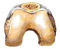
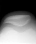
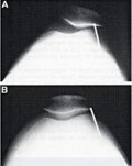
Neutral position
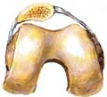
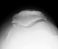
Subluxated or tilted
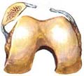
Full dislocation
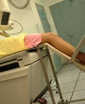
Xray Position
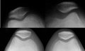
thick cartilage space
thin space and bone spurs
tilted alignments on top
lateral tilting
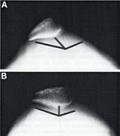
before / after lateral release

at risk for slipping out
/ subluxating
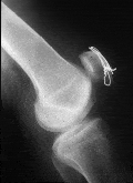
Fixed with a wire
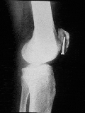
Fixed with screws
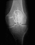
Fixed with Pins and wire
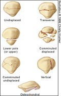
Patella Fracture Patterns
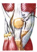
lateral release medial repair
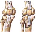
tibial tubercle transfer